
Medical Testimony on the Headwound
Did the Parkland Doctors See Cerebellum?
Most conspiracists believe the large gaping wound on Kennedy's head was in the rear in occipital bone. This supposedly proves that a shot from the Grassy Knoll hit Kennedy from the right front and blew out the back of the head. One key piece of evidence they offer is the fact that several Dallas doctors said they saw cerebellar tissue in Kennedy's head wound.
The cerebellum is nestled beneath the parietal, temporal, and occipital lobes of the brain (see drawing at right, above). If it really was badly lacerated with pieces oozing from the wound, this would certainly imply damage low on the back of the head.
The doctors' statements seem clear enough on this issue.
- McClelland: ". . . you could actually look down into the skull cavity itself and see that probably a third or so, at least, of the brain tissue, posterior cerebral tissue and some of the cerebellar tissue had been blasted out. . ." 6H33
- Carrico: "One could see both cerebellum and cerebrum fragments in that
wound." 7 HSCA 268
- Clark: "Both cerebral and cerebellar tissue were extruding from the
wound." 17H9-10, CE-392
- Jenkins: "...even to the extent that the cerebellum had protruded
from the wound." 17H14-15, CE-392
- Baxter: "....with this and the observation that the cerebellum was
present...." 6H41
- Perry: "...and there was visible brain tissue in the macard and some
cerebellum seen." 7HSCA302
- Peters: "You could directly into the cranial vault and see cerebral injury to the cerebral cortex and I thought
at the time to the cerebellum." ARRB interview with Parkland Hospital doctors, 8-27-98, p. 52
- Grossman (D. Horne's words): ". . . one [wound] was a circular puncture in the occipital region . . . 2 cm. in diameter . . . through which he could see brain tissue which he believes was cerebellum. . . ."
ARRB, MD# 185
The uniformity of their testimony is impressive, and unlike the testimony on the location of the wound, the doctors' various statements over the years have been consistent in saying "cerebellum" (Gary Aguilar, "The Converging Medical Case for Conspiracy in the Death of JFK," in Murder in Dealey Plaza, pp. 175-218).
But it's also the case that eyewitness testimony is inherently suspect. Indeed, there are cases where multiple corroborating witness are clearly wrong, as was the case with the "Alice witnesses," and the Dealey Plaza witnesses who said they saw a motorcycle officer drive his bike part way up the Grassy Knoll. But what about this case?
Is there better evidence than the Parkland witnesses? Yes. First, there are the autopsists, who didn't just glance at the head wound, but who actually removed the brain from the head and examined it. What did they say?
The Assassination Records Review Board was quite aware of the issue, and pressed both Humes and Boswell about it. First, Jeremy Gunn of the ARRB interviewing Boswell:
Q. Do you recall whether it was particularly easy to remove the brain?
A. I think it was a routine procedure. In Dallas, they had said that the cerebellum was the part of the brain that was injured and exuding. But they were wrong because the cerebellum is enclosed in a dural sort of compartment, and in order to get the cerebellum out, you have to cut the dura around, and then you—that's the only hard part about getting the brain out. And the manner in which we were doing it, both the cerebral hemispheres were already exposed without dura, and it was really very simple to take out.
Q. During the course of the autopsy, did you have an opportunity to examine the cerebellum?
A. Yes.
Q. And was there any damage to the cerebellum that you noticed during the time of the autopsy?
A. No.
Q. So both the right and left hemisphere of the cerebellum were intact?
A. Yes.
Q. Was the tentorium damaged at all?
A. No.
Humes testimony is a bit more complicated, since he says the cerebellum was "somewhat disrupted," but believes that was the result of the pressure of the bullet passing through the skull. He testified to no lacerations of the cerebellum.
So it seems that the autopsists, who removed the intact cerebellum from the skull, don't agree with the Dallas doctors about the damage.
But is there evidence that's better than the autopsists testimony? Most certainly. Some of the photos of the brain, taken at the autopsy, show the inferior aspect. These photos were examined by the House Select Committee's Forensic Pathology Panel. They described what they show as follows. Be warned that the description is a bit tedious, but shows the level of detail they could see in the photos.
(330.) ... Color transparencies and prints Nos. 46,47, 48, and 49 and
black and white prints Nos. 19, 21, and 22 reveal the inferior aspect
of the brain, with extensive fragmentation and laceration of the right
inferior cerebral hemisphere, some loss of cerebral substance on the
inferior surface of the left temporal lobe, and scattered areas of
subarachnoid hemorrhage in the underlying cortex. The right sylvian
fissure shows dark red-brown to black discoloration suggestive of
blood clot. The surface of the midtemporal region is lacerated and
depressed. The cerebral peduncles are likewise lacerated. The panel
notes that the posterior-inferior portion of the cerebellum virtually
intact. It certainly does not demonstrate the degree of laceration,
fragmentation, or contusion (as appears subsequently on the superior
aspect of the brain) that would be expected in this location if the
bullet wound of entrance were as described in the autopsy report [in occipital bone].
(House Select Committee on Assassinations, Volume 7, p. 129)
In testimony before the Committee, Michael Baden, Chairman of the Panel, explained as follows:
Dr. BADEN - . . . We, as the panel members, do feel after close examination of the negatives and photographs under magnification of that higher perforation, that it is unquestionably a perforation of entrance; and we feel very strongly, and this is unanimous, all nine members, that X-rays clearly show the entrance perforation in the skull to be immediately beneath this perforation in the upper scalp skin; and further, although the original examination of the brain was not complete, photographs of the brain were examined by the panel members, and do show the injury to the brain itself is on the top portion of the brain. The bottom portion or undersurface of the brain, which would have had to have been injured if the bullet perforated in the lower area as indicated in the autopsy report, was intact. If a bullet entered in this lower area, the cerebellum portion of the brain would have had to be injured and it was not injured.
(House Select Committee on Assassinations, Volume 1, p. 301, emphasis added)
Note that neither of these statements addresses the question of whether the back of Kennedy's head was blown out — which was an absurd notion given what the photos and x-rays show. Rather, the issue was whether the inshoot was low near the EOP (as the autopsy doctors placed it) or higher in the cowlick area (where the photos and x-rays show it). But the observations about the cerebellum stand.
Just what was going on here? Was this some kind of mass halucination or something?
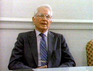 In 1988 Public Broadcasting's NOVA talked to four of the Parkland doctors and allowed them to examine the autopsy photos and x-rays in the National Archives in Washington. Marion "Pepper" Jenkins (right), one of the witnesses who said he saw cerebellum, explained:
I did say cerebellum in my first official report. And the cerebellum ordinarily is in a posterior part. And here I know very well that the wound was more anterior than that, but there was a portion of the brain that looked like it had a stalk, and is convoluted to look like what I thought was cerebellum.
Jenkins' colleague Paul Peters also viewed the materials in the Archives, and told NOVA:
I said that I thought perhaps part of the cerebellum was missing, and that shows how even a trained observer can make an error in moment of urgency.
Thus there was something in the bloody mass of blood and tissue that looked like cerebellum. But it wasn't cerebellum. The doctors weren't crackpot witnesses, they were just wrong.
Thanks to John Canal and Jerry McNally for bringing key sources used on this page to the author's attention.
 Return
to Kennedy Assassination Home Page
Return
to Kennedy Assassination Home Page