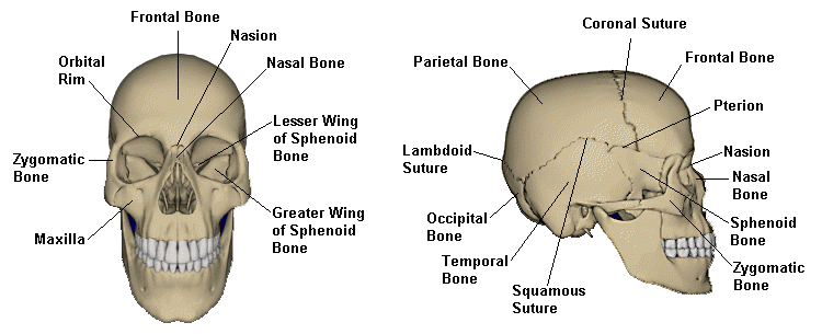
I assembled a list of features noted by the HSCA radiologists in their reports. I numbered the list and located the features on the X-rays to the best of my ability. At the bottom of the page are labeled frontal and lateral skull models for reference.

The following initials indicate the source of the quote:
| DD | Dr. David O. Davis | |||||||||||
| GM | Dr. G.M. McDonnel | |||||||||||
| RR | RADIOLOGY review of a Randy Robertson manuscript. | |||||||||||
| 1. | GM: Nearly complete loss of right parietal bone, the upper portion of the right temporal bone, and a portion of the posterior aspect of the right frontal bone. DD: .there is apparently absence of bone anterior to this line, with the absence present to a point approximately equivalent to where the coronal suture on the right side should be. |
| 2a. | GM: A metallic fragment on the outer table of the right occipital bone 9.6 cm. above the mid portion of the external occipital protuberance (EOP). DD: There is a metallic fragment about 9 or 10cm above the external occipital protuberance, which metallic fragment is apparently imbedded in the outer table of the skull. On the frontal view, this metallic fragment is located 2.5cm to the right of midline, and on the lateral view, it is approximately 3-4cm above the lambda. |
| 2b. | GM: 1 cm. above the metallic fragment is a depressed fracture from which stellate type fractures "radiate" into both occipital bones, the right parietal bone and the right temporal bone. |
| 3. | DD: There are a large number of fractures in the calvarium, and the linear fractures seem to more or less emanate from the imbedded metallic fragment, and radiate in a stellate fashion in various directions. |
| 3a. | DD: There is a large fracture extending directly anteriorly along the sagittal suture, which is, at least at the point visualized, widely separated. This fracture seems to extend into the frontal bone, more or less at the midline, down to the frontal sinus which is also fractured. GM: There are fracture lines through the anterior and posterior aspects of the right frontal sinus with air in this sinus. There is a metallic fragment above the sinus appearing to be between the bony tables of the frontal bone. |
| 3b. | DD: There is a sharply defined linear fracture extending laterally from the metallic fragment into the left side of the calvarium, around the parietal bone to the lateral aspect of the skull. |
| 3c. | DD: Two linear fractures extend inferolaterally from the metallic fragment, one into the occipital bone, about 3cm from the midline, and this fracture crosses the lambdoid suture. |
| 3d. | DD: The other one is more lateral, and extends down toward the lateral sinus, probably above the lambdoid suture. |
| 3e. | DD: Additionally, there is a fracture line extending more or less laterally from the metallic fragment toward the temporal bone on the right side, which is identified only by the anterior edge of the posterior fragment, since there is apparently absence of bone anterior to this line, with the absence present to a point approximately equivalent to where the coronal suture on the right side should be. |
| 3f. | DD: I neglected to describe in the text of this report an extensive fracture which extends inferolaterally from the impact point toward the left side which probably reaches the temporal bone or at least the mastoid region after crossing a goodly portion of the occipital bone there. |
| 4. | DD: There is a fracture fragment inferior to the absent bone, with the corner of the fragment extending down to the parietal squamosal suture, and this fragment is displaced from its normal position as indicated by overlap of the infero and posterior aspects of the fracture fragment. |
| 5. | GM: There is a fracture line extending through the floor of the sella turcica with bony fragments in the sphenoid sinus. |
| 6a. | GM: A linear alignment of tiny metallic fragments is associated with the entry, path travel, and exit in the posterior aspect of the right frontal bone. |
| 6b. | DD: There are a number of metallic fragments extending anteriorly from the inner table of the skull at a point approximately 6cm antero-superiorly from the previously described imbedded metallic fragment. These fragments extend inferoanteriorly across the entire skull and actually project (on other images that I have seen) in a fashion that suggests that the the large fragment is outside the intracranial space. Presumably this represents a metallic fragment in the scalp, although this cannot be accurately determined from this particular examination. |
| 7. | DD: It also seems reasonable to assume that the exit point is near the coronal suture on the right side, about 5 or 6, or perhaps slightly more, cm above the pterion. |
| 8. | RR: This fracture pattern is discussed in a paper by Smith, et al. In the JOURNAL OF FORENSIC SCIENCES 1987.in which they explain how acutely increased internal pressure during a projectile injury produces concentric heaving fractures. |
Other Features Noted but not Marked
DD: The right orbital rim is also fractured laterally, and the roof of the orbit is fractured on the right side, as is the inferior orbital rim, indicating that there is an unstable orbit.
GM: There is elevation of the galea medial and lateral, as well as anteriorly, to the depressed fracture in the right occipital bone. A small metallic fragment lies medial to the fracture site between the galea and the outer table of the skull.
GM: There are fracture lines through the anterior and posterior aspects of the anterior ethmoid cells with air in the right side anterior ethmoid.
| Back to Reading the X-rays | Contents | Continue to The Dox Drawing |