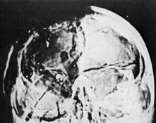
The area from the right orbit (eye socket) to the top of the head appears dark in the non-enhanced AP X-ray (above). A common assumption is that this must be all missing bone. But, what at first seems to be the lower margin of a skull defect is nothing more than a natural feature in the skull, visible on most all frontal skull X-rays.
Harry Livingstone published these two drawings in this book, High Treason 2.
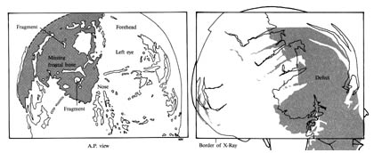
He claims the X-ray indicates bone is missing in the facial skeleton. Since the autopsy photos show no such damage, he concludes the X-rays are forgeries.
Dr. John Lattimer viewed the original X-rays and drew these two sketches:
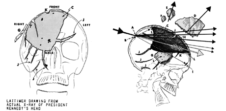
He says that he did not intend the gray region in the AP to represent missing bone, that it is just a diagram of how the X-ray appears. However, comparing the fractures labeled "J" to those in the lateral sketch at the right indicates the gray area in the AP is supposed to be the same defect as the one seen in the lateral. It would seem, then, that Lattimer initially interpreted the dark area within and above the right orbit to be missing bone just as Livingstone does, but in the parietal-frontal region rather than the face.
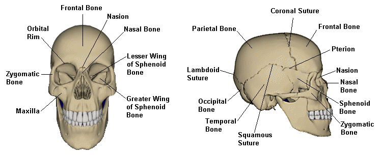
The Sphenoid Bone
Both of these sketches of the AP X-ray misidentify the lesser wing of the sphenoid bone as the lower edge of the defect. The sphenoid bone is visible within the orbits in the frontal view of the skull:
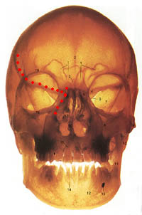
One difference between this image and the AP X-ray is that this image is a straight frontal view rather than the Modified-Waters projection of the AP. To achieve better alignment, the skull should be tipped back which will move the sphenoid lower in the orbits.
In the right image, I drew red dots marking the bottom and lower left edge of the skull defect as drawn in the Livingstone and Lattimer sketches. (The line ends don't meet because this is a frontal view, which positions the sphenoid higher.)
The sphenoid is also clearly visible in the left orbit on the AP X-ray.
Why the Apparent Skull Defect is only Apparent
The black region in the non-enhanced AP X-ray seems to be missing bone because:
- The edge of the lessor wing of the sphenoid and the edge of the nose are natural contours that the eye is drawn to.
- The orbits are naturally darker than the regions around them. The right orbit is darker because brain matter is missing and/or air is present in the tissues. HSCA radiologist G.M. McDonnel noted "subdural air over the left parietal hemisphere." (The upper edge of the occipital bone is near the bottom of the orbits in the AP X-ray.) This darkens the general area. Also notice the right sphenoid is a bit darker than the left one, meaning the region of missing tissue and/or air extends down to at least the lower orbital rim.
- The contrast range of the X-ray is much greater than what can be reproduced in print. The darkening of the orbital region pushes it below the threshold where it will be printable. It prints as a solid black. The sphenoid bone is dense enough that it is not darkened enough to drop to black.
- The area above the right orbital rim represents intact frontal bone, some missing brain matter, and a large section of missing parietal bone. The frontal bone by itself is not dense enough to produce a bright enough image to be visible. And as you can see on the intact left side of the forehead, the skull naturally darkens towards the lateral edge.
Thus, in the non-enhanced AP X-ray, the combination of the factors listed above cause the orbital and supra-orbital regions to reproduce as one large black area with well-defined edges. The edges, however, are merely the natural contours of the lesser wing of the sphenoid bone and the nose.
The enhanced AP X-ray, below, shows large fractures within the parietal and occipital bones at the back of the skull. Fracture lines cannot form in missing bone. The right side of occipital bone must be there. And since the upper rim of the right orbit is visible, frontal bone must be there. Both the rear and the face of the skull are intact, albeit, fractured to varying extents. The only region that is not visible, even after enhancement, is the right parietal, or upper rear region, of the skull.