The Thoracic Spine
 
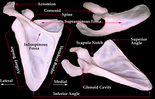
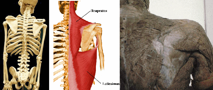
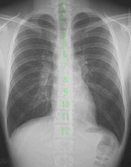
Each of the 12 thoracic vertebrae are attached to a pair of ribs. This part of the skeleton houses almost all of the vital organs. The ribs serve as a bony cage for protection of the delicate organs. Unlike the cervical spine, the thoracic spine is flanked on both rear sides by additional bones that provide protection. The rear sides have additional protection via the scapulae, or shoulder blades. These are thick, dense bones that serve as additional protection as well as an attachment for many muscles. The attachment of muscles provide leverage points for muscular contraction which allows us to move our arms freely. Without the shoulder blades, we would have very limited motion of the arms because muscles such as the trapezius, teres minor, teres major, supraspinatus, infraspinatus, levator scapula, latissimus dorsi, serratus anterior, rhomboids and others would not have complete attachments and could not contract fully.  
 
The thorax, or the area between the first and twelfth thoracic vertebrae, consists of 24 ribs, 2 clavicles, 2 scapula, 12 vertebrae and the sternum. Of all of these bones, the scapula has the greatest freedom of movement. It's only articulation is at the shoulder where it forms the glenohumeral joint and the acromioclavicular joint. Since the shoulder does not form a joint near the middle, it has less natural restriction in movement. Therefore, the position of the scapula can vary greatly. However, we can assess some common reference points with a person in a neutral, relaxed position.  


Scapular Anatomy and Landmarks
 
The scapula is a triangular shaped bone on the rear portion of the thorax. It overlies the rib cage and covers parts of the 2nd to 7th ribs. It connects the clavicle, or collarbone, to the upper arm. This forms the shoulder girdle. The shoulder joint has the greatest freedom of motion of any joint in the human body. This freedom of movement comes at the expense of the joint's laxity and instability.  
 
In an anatomically neutral position, portions of the scapula can be related to other bony parts of the body. The superior angle of the scapula is commonly found at the level of the 2nd thoracic vertebra. The inferior angle of the scapula is located adjacent to the spinous process of the seventh thoracic vertebra (Clinically Oriented Anatomy, 3rd Ed., Keith L. Moore) The acromion process is commonly located within the T1-T2 level (Atlas of Human Anatomy, Frank M. Netter, MD). This bony prominence can vary position depending upon the level of muscular contraction of the trapezius and levator scapula. If minimally contracted, the acromion may be found slightly higher than T1. You can see this by shrugging your own shoulders.
 
 
The Human Skeleton vs. External Appearance
 
 
Compare these illustrations. It should become apparent that the addition of the musculature to the spine changes it's outward appearance significantly. The trapezius muscle is the largest muscle of the upper back and contributes greatly to the appearance. Specifically, the lateral (meaning away from the center) portion attaches to the shoulder blade. The fibers run diagonally upwards and attach to the spine and at the external occipital protuberance (EOP) . This forms an upward slope, obscuring the true cervicothoracic junction (marked at right). This 'masking' effect has led many researchers and casual readers to assume that the autopsy photo shows an 'obvious' misrepresentation. This misconception allows many ill-informed readers to conclude a conspiracy on the basis of an erroneous assumption. The last photograph is from a cadaver with the skin removed to show the fibers of the trapezius and the overall contour of the upper back.